Cell-based assays are the main stay in drug discovery projects, where collections of compounds are screened to assess effects on cell viability, cell proliferation or show direct cytotoxic effects that eventually lead to cell death. Assessing cell viability is a key step in daily cell manipulation and often a requirement for subsequent processing and analysis. Cell proliferation and the characterization of chemicals that either promote or inhibit cell proliferation are important areas of cell biology and drug discovery research.
Enzo Life Sciences offers a comprehensive product portfolio for
Live Cell Analysis. Our CELLESTIAL® assay platform offers fluorescence-based live cell assays that are sensitive, highly specific, and optimized for the most demanding imaging applications, including microplate-based high content screening, flow cytometry, confocal microscopy, and wide-field fluorescence microscopy where consistency and reproducibility are essential.
Cell Viability
There is a wide range of cell viability methods available.
ApoSENSOR™ Cell Viability Assay utilizes bioluminescent detection of the ATP level for a rapid screening of apoptosis and cell proliferation simultaneously in mammalian cells. The assay utilizes luciferase to catalyze the formation of light from ATP and luciferin, and the light can be measured using a luminometer or Beta Counter. The ATP assay is the fastest cell viability assay to use, the most sensitive, and is less prone to artifacts than other viability assay methods.
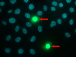
Distinguishing between live and dead cells is very important for investigation of growth control and cell death. The
Live-Dead Cell Staining Kit provides ready-to-use staining reagents to conveniently discriminate between live and dead cells. The kit utilizes Live-Dye™, a cell-permeable green fluorescent dye (Ex/Em = 488/518nm), to stain live cells. Dead cells can be easily stained by propidium iodide (PI), a cell non-permeable red fluorescent dye (Ex/Em = 488/515nm).

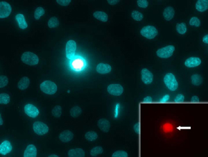
Figure 1. NUCLEAR-ID® Red/Green dye is detected as red-stained nuclei in live cells and fluorescent-green nuclei in dead cells (inset, arrow).
|
is a mixture of a red fluorescent cell-permeable nucleic acid dye and a green fluorescent cell-impermeable nucleic acid dye that is suited for staining dead nuclei. The staining pattern arising from the simultaneous combination of these two dyes permits determination of live and dead cell populations by fluorescence/confocal microscopy.
Cell Proliferation
Reagents for counting cells and quantitating cell proliferation are valuable research tools.
Cell Counting Kit-8 provides a sensitive colorimetric assay for the determination of cell viability in cell proliferation and cytotoxicity assays. The assay utilizes a highly water-soluble tetrazolium salt, WST-8, which is reduced by dehydrogenase activities in live cells to give a yellow-color formazan dye. WST-8 is more stable and less cytotoxic compared to other tetrazolium salts, making it especially useful for longer incubation periods.The amount of the formazan dye, generated by the activities of dehydrogenases in cells, is directly proportional to the number of living cells. The detection sensitivity of CCK-8 is higher than the other tetrazolium salts such as MTT, XTT, MTS or WST-1 as shown in Figure 4. Treated and control cells can be differentiated in this cell proliferation assay to distinguish cytotoxic effects. This is a homogeneous cell viability assay developed for a 96-well format that is suitable for high throughput screening.
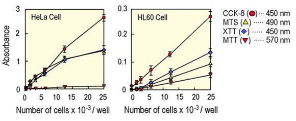
Figure 4. Comparative data of Cell Proliferation Assay, CCK-8
|
Cell tracking assays have the common goal of determining the fate of a particular cell population within a heterogeneous environment, whether
in vivo or
in vitro. The
CYTO-ID® Green and
CYTO-ID® Red Long Term Tracer kits are suitable for tracing cell lineages, as well as assaying proliferation, chemotaxis, migration, phagocytosis, and cell- and antibody-mediated cytotoxicity. Analysis of labeled and unlabeled cell populations over time by flow cytometry or microscopy is also feasible. Long-term fluorescent tracing of live cells, especially different cell populations, has been difficult due to the lack of suitable fluorescent probes. These probes incorporate into cells through non-covalent, hydrophobic interaction with the lipid hydrophobic portion of the cell membrane. As many as four cell populations can be monitored simultaneously through incorporating green, red or mixtures of green and red dyes into the plasma membranes of cells. These dyes are well-retained for 96-120 hours and faithfully transferred to daughter cells upon mitosis, without displaying a tendency for nonspecific transfer to adjacent unlabeled cell populations. Normal physiological functions of cells are well preserved since these dyes do not covalently modify any vital biomolecules in the cell or disturb their functions. These new dyes are powerful probes for analyzing long-term cell viability, cytotoxicity, cell adhesion, cell migration, lineage tracing and cell-cell fusion. Cells labeled with these probes can be analyzed with flow cytometry and fluorescence microscopy.
Cytotoxicity
Cytotoxicity, or the quality of being toxic to cells, is a cellular regulator which can result in a variety of cell fates. Most often, it results in different types of cell death including necrosis, apoptosis and other fates. Enzo provides a variety of assays to quantify cytotoxicity in cells, including an LDH Cytotoxicity WST Assay. The
LDH Cytotoxicity WST Assay is a colorimetric assay kit used to determine cytotoxicity by measuring lactate dehydrogenase activity released from damaged cells. Lactate dehydrogenase (LDH) is a stable enzyme, present in all cell types, which is rapidly released into the cell culture medium upon damage of the plasma membrane. LDH is the most widely used marker in in cytotoxicity studies. Assays that measure metabolic activity are suitable for analyzing proliferation, viability, and cytotoxicity
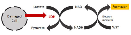
Figure 5. Mechanism for LDH Cytotoxicity WST Assay
|
The progression of the cell cycle is controlled by a complex interplay among various cell cycle regulators that either stimulate or inhibit the cell from entering each stage of the cell cycle. Dysfunction of any step in this regulatory cascade causes abnormal cell proliferation which underlies many human pathological conditions, such as cancer.
NUCLEAR-ID® Green and
NUCLEAR-ID® DNA stain are novel DNA-intercalating fluorescent probes that freely enter live cells, allowing cell cycle analysis without laborious permeabilization and fixation steps. Since the dye intercalates exclusively into double-stranded DNA, no RNase treatment is required and the fluorescence intensity of stained cells is directly proportional to their DNA content.
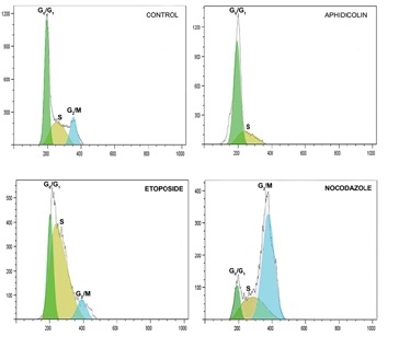
Figure 6. Drug treatments with live cells that inhibit cell cycle progression at different phases.
|
Cell-based assays for evaluating the functional status of mitochondria are emerging as useful tools for elucidating the role of mitochondrial activity in drug-induced toxicity.
MITO-ID® Membrane Potential Detection Kit measures mitochondrial membrane potential with a cationic dye that fluoresces either green or orange depending on membrane potential status. In energized cells, the MITO-ID® Membrane Potential reagent exists as a green fluorescent monomer in the cytosol and also accumulates as orange-fluorescent aggregates in the mitochondria. However, in cells with compromised mitochondrial membrane potential, the MITO-ID® Membrane Potential reagent exists primarily as green-fluorescent monomers throughout the cytosol and no longer exhibits orange fluorescence in the mitochondria. The
MITO-ID® Membrane Potential Cytotoxicity Kit measures fluctuations in mitochondrial membrane potential (MMP) utilizing a cationic dual-emission dye that exists as green fluorescent monomers in the cytosol, and accumulates as orange fluorescent J-aggregates in the mitochondria. Mitochondria having a low membrane potential will accumulate low concentrations of dye and will exhibit green fluorescence while more highly polarized mitochondria will exhibit orange-red fluorescence. Cells exhibit a shift from orange to green fluorescence as mitochondrial function becomes increasingly compromised.
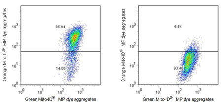
Figure 7. Flow cytometric analysis of control and treated cells. Untreated Jurkat cells (left) and were treated with 1 μM CCCP for 15 mins (right). Cells were then stained with Enzo MITO-ID® Membrane Potential Dye and run on a FACS Calibur instrument.
|
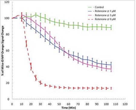
Figure 8. Real-time detection of mitotoxicity in drug screening. Time-course study of mitochondrial membrane potential changes using a BioTek Synergy™ Mx fluorescence microplate reader. HeLa cells were incubated with MITO-ID® MP dye for 30 minutes at room temperature (no serum or media removal). Rotenone was added to achieve concentrations of 1 µM, 3 µM and 9 µM. MITO-ID® MP dye was shown to be responsive to rotenone, as demonstrated by a decrease in orange signal.
|
Enzo Life Sciences offers a range of products for your
Cellular Analysis research needs. As Scientists Enabling Scientists, we realize the value in providing relevant information to our customers working in the fields of life sciences, drug development and clinical research. We are happy to provide simple yet useful tips and guidance for your research needs. Please check out our
successful research tips or contact our
Technical Support Team for further assistance.